 ACDS - Automatic Cell Detection and Segmentation
ACDS - Automatic Cell Detection and Segmentation
ACDS - Automatic Cell Detection and Segmentation
Our proposed method to detect and segment the phytoplankton cells from microscopic images of non-setae species.
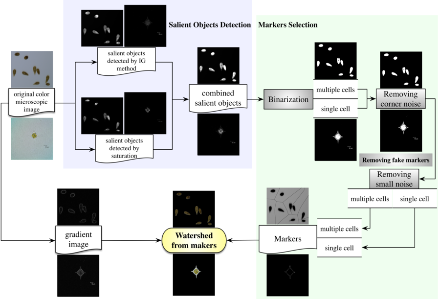
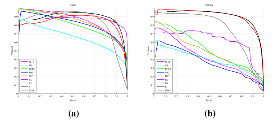
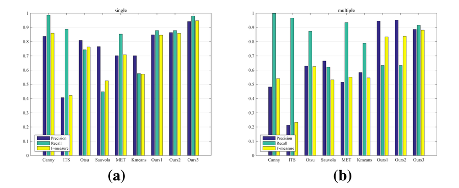
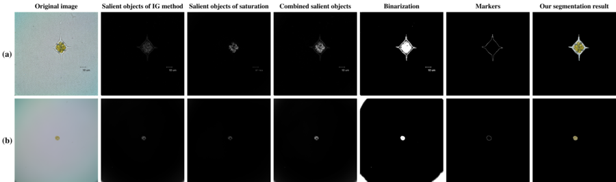
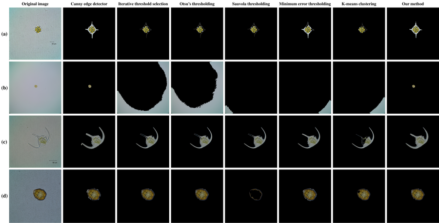
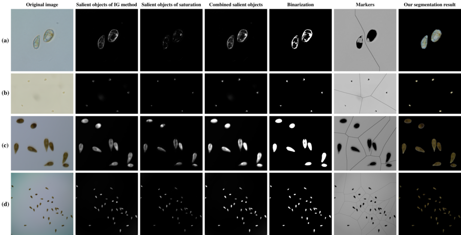
Saliency-based marker-controlled watershed method was proposed to detect and segment phytoplankton cells from microscopic images of non-setae species. This method first improved IG saliency detection method by combining saturation feature with color and luminance feature to detect cells from microscopic images uniformly and then produced effective internal and external markers by removing various specific noises in microscopic images for efficient performance of watershed segmentation automatically. We built the first benchmark dataset for cell detection and segmentation, including 240 microscopic images across multiple phytoplankton species with pixel-wise cell regions labeled by a taxonomist, to evaluate our method. We compared our cell detection method with seven popular saliency detection methods and our cell segmentation method with six commonly used segmentation methods. The quantitative comparison validates that our method performs better on cell detection in terms of robustness and uniformity and cell segmentation in terms of accuracy and completeness. The qualitative results show that our improved saliency detection method can detect and highlight all cells, and the following marker selection scheme can remove the corner noise caused by illumination, the small noise caused by specks, and debris, as well as deal with blurred edges.
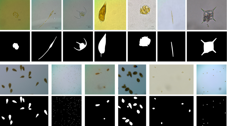
We construct a new dataset that contains 240 phytoplankton microscopic images with 225 of single cell while 15 of multiple cells and human labeled ground-truth cell regions. These images are acquired and selected with the help of phytoplankton experts in different sizes from $256\times 256$ to $4080\times 3072$ and different species of non-setae phytoplankton. The pixel-wise ground truth masks are produced guided by phytoplankton taxonomist to contain the biomorphic characteristics of cells. These data provide useful resource to study the phytoplankton for automatic detection and segmentation. Some sample images are shown in the above figure. The data are available for downloading here.
The MATLAB source code can be followed and downloaded from https://github.com/zhenglab/ACDS.
| No. | Canny | ITS | Otsu | Sauvola | MET | Kmeans | Ours1 | Ours2 | Ours3 | GT |
|---|---|---|---|---|---|---|---|---|---|---|
| #1 | 10 | 55 | 11 | 51 | 12 | 12 | 16 | 14 | 12 | 12 |
| #2 | 8 | 7 | 7 | 57 | 7 | 8 | 10 | 10 | 9 | 9 |
| #3 | 158 | 839 | 745 | 203 | 177 | 455 | 267 | 198 | 125 | 141 |
| #4 | 207 | 3390 | 12 | 11 | 46 | 9 | 9 | 5 | 3 | 3 |
| #5 | 12 | 5323 | 6 | 46292 | 44 | 14232 | 8 | 2 | 2 | 2 |
| #6 | 35 | 107 | 6 | 5220 | 49 | 3014 | 17 | 2 | 2 | 2 |
| #7 | 10 | 228 | 10 | 14 | 27 | 10 | 11 | 9 | 5 | 5 |
| #8 | 8 | 11 | 5 | 4 | 11 | 3 | 21 | 3 | 2 | 2 |
| #9 | 35 | 345 | 338 | 53 | 32 | 34 | 39 | 35 | 31 | 31 |
| #10 | 6 | 614 | 9 | 7 | 9 | 4 | 3 | 2 | 2 | 2 |
| #11 | 18 | 627 | 336 | 90 | 58 | 114 | 32 | 14 | 12 | 11 |
| #12 | 35 | 224 | 37 | 42 | 121 | 33 | 30 | 29 | 29 | 31 |
| #13 | 8 | 390 | 8 | 10 | 24 | 7 | 13 | 10 | 5 | 5 |
| #14 | 11 | 11 | 11 | 26 | 11 | 11 | 12 | 11 | 10 | 11 |
| #15 | 12 | 6934 | 4 | 41825 | 114 | 15283 | 47 | 21 | 5 | 2 |
We wish to thank the Algal Collection of Research Center for Harmful Algae and Aquatic Environment in Jinan University and Key Laboratory of Marine Environment and Ecology, Ministry of Education in Ocean University of China for providing the samples of phytoplankton species and the instruments to observe and acquire the corresponding microscopic images. This work was supported by the National Natural Science Foundation of China under grant numbers 61301240 and 61271406 and China Postdoctoral Science Foundation under grant number 2016M590658.

